I generally turn in the content for Crystallography Times a week or so before it is sent out. This gives Bev Vincent and Amanda Cochran-Kelly plenty of time to fix the problems and generate the content you receive in your inbox. Kudos to the two of them for supporting Crystallography Times. The time delay is why I am going to talk about something that happened nine days ago in the next paragraph. | | The eclipse of 2024, as seen from Temple, TX. Photo courtesy of Chris Reyes. | Yesterday, a swath of North America experienced a total solar eclipse. It is good that I got to see the total eclipse in New York City in March of 1970, as Houston weather did not cooperate one iota yesterday. Those of you who were in or drove to the path of totality, I hope you had good weather.
This month, we interview Chris Malliakas of Northwestern University, the first recipient of a XtaLAB Synergy-ED in the US. We also introduce members of the application laboratory at our office in Frankfurt, Germany. Khai-Nghi Truong provides the tip of the month on the use of Proffitloop. Jeanette reviews Her Space, Her Time: How Trailblazing Women Scientists Decoded the Hidden Universe by Shohini Ghose.
I wasn’t sure I would raise the issue of the deaths last week of the seven World Central Kitchen aid workers, but there was a request for support from WCK in my personal inbox last night. WCK is a great NGO that provides food to those in need around the world who are in dire straits. Here is a link to provide support.
Be safe,
Joe Ferrara | | | | | | Explore the diverse realm of sub-micron crystal structures through the lens of the XtaLAB Synergy-ED. This webinar delves into its applications across a wide spectrum of materials, including small molecules, MOFs, COFs, biological samples, minerals, and beyond. Highly beam-sensitive samples or measured under cryogenic conditions, the XtaLAB Synergy-ED offers exceptional capabilities for precise analysis. Discover how this instrument contributes to advancements in chemistry, materials science, and related fields.
Wednesday, May 29, 2024 at 16:00 CET Time Zone Converter >
Register Now > | | | | Interview with a Crystallographer Christos Malliakas, Northwestern University | | | When did you start in structural science and how long have you been using microED in your research? It was my first semester as a graduate student, so fall 2002, when I started using both X-ray and electron diffraction for my thesis work. At that time, I was not using the term microED or 3D-ED of course. There were different terms, but most of the time it was called “selected area electron diffraction on single crystals.”
For the X-ray portion of my work, I started using powder and single crystal diffractometers before having an official class because I learned how to operate them from lab-group members. This was not the case for the electron microscopy / diffraction work, for which I had to take formal classes before I could use the instruments.
What first attracted you to microED? Having access to a very strong beam and being able to get information from not only very small in size features but also very weak features in diffraction was the main reason why I started using electrons. For example, I was studying the formation of supercells in extended structures and part of the benefit of electron diffraction (at that time) was the detection of very weak reflections along with very strong reflection on an analog medium such as phosphorus screen or negative photographic film or image plate.
What is the biggest challenge you are facing in your research just now? One of the big challenges is the structure elucidation of large numbers of samples generated by high-throughput syntheses using robots. What is needed here is a highly optimized workflow and protocol to perform very fast initial screening of small crystallites. This is mainly needed for answering fundamental questions, such as: did we make something new? Did we make something interesting? After a successful rapid initial screening, identification of candidates and more efforts on crystal growth and more conventional synthesis could follow. The main problem right now is essentially not only complexity of samples, but also the large number of samples that is coming out from all these combinatorial high throughput syntheses. We already have tools and methods for high-throughput powder analysis but what is missing is the 3D analogue for full structure elucidation.
How does microED fit into your main research interest? Structure elucidation of very small size crystallites is the main interest. microED enables the 3-dimensional structural characterization of very small crystallites that are in the sub-micron to nanometer size range. microED bridges the size gap between the minimum size crystals which can be used in X-ray single crystal diffraction experiments (with either inhouse or synchrotron sources) and the typical sizes needed for electron diffraction measurements. This is critical for the high throughput projects that tend to generate small size crystals. Furthermore, projects that yield small crystals that are difficult to grow, for example natural synthesis, can also highly benefit from microED. Getting 3-dimensional structural information from materials that are at the early stages of development is very important especially during the screening process for finding good candidate materials for a given application.
Looking into your crystal ball, how do you think microED will change in the next 10 years? I believe the introduction of established techniques and hardware components already used in standard electron microscopy/diffraction is the natural step in near-future developments. For example, the use of energy filters might be easy to implement to suppress inelastic scattering or even to perform energy/element specific scattering experiments.
I think the other advantage an electron diffractometer can have (as opposed to in-house X-ray diffractometers) is the potential of combining elemental analysis by having an EDS detector on the electron diffractometer, so one can perform diffraction and elemental analysis on the same grain.
Additionally, pump-probe ultrafast electron diffraction at femtosecond timescales might also be commercially available with microED measurements. Non-microED versions of this type of instrumentation do exist that can perform pump-probe measurements by using lasers and electrons at the same time. There are a few research groups out there that do this type of in-house ultra-fast electron diffraction measurements using not necessarily EMs but a combination of high vacuum chambers, e-guns, fast detectors, etc.
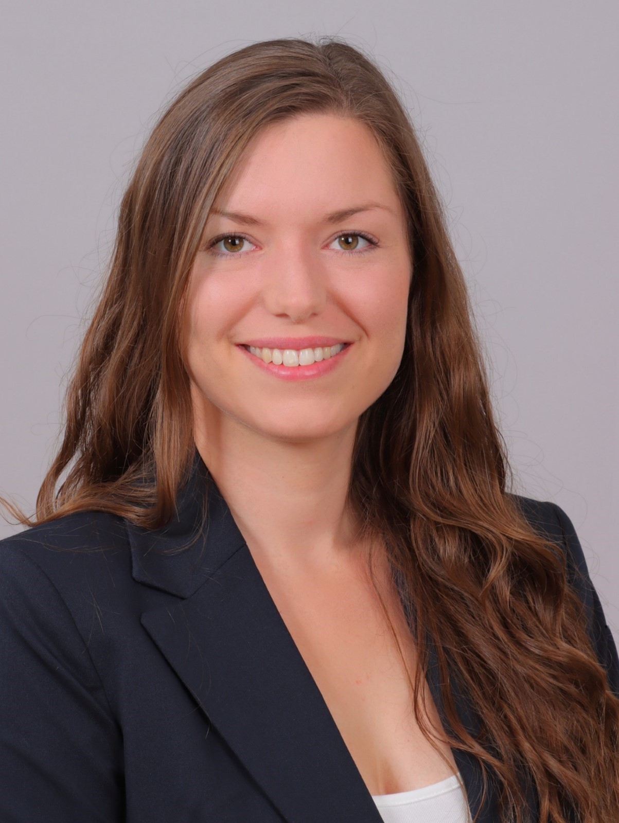
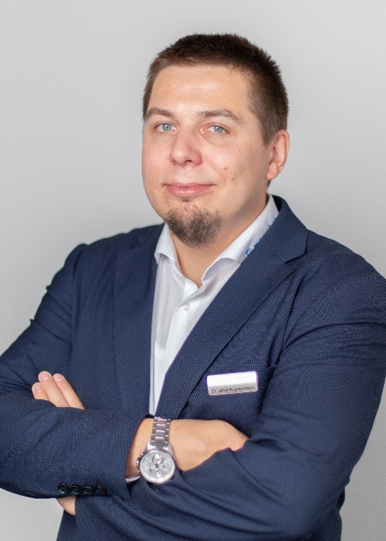
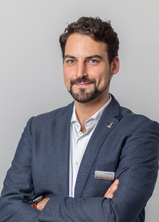
Another interesting area is total scattering experiments using electron diffraction. This requires a better quantitative approach where both diffuse intensities and Bragg reflections are used properly in order to extract information about the sort-, medium, and long-range ordering in a sample. Success of quantitative total scattering and even precise 3D diffraction analyses will depend on the advancement of software and theory that takes dynamical effects into account. | | | | | Our European application lab is conveniently located 15 minutes from Frankfurt International Airport and hosts five application scientists dedicated to single crystal diffraction. We handle all sorts of samples and use many different techniques to analyze single crystals and micro powders. | | Rigaku's European application lab | Our application team members were trained as chemists, all with strong backgrounds in single crystal diffraction of various types. Measurements of crystals smaller than one micrometer require the use of electron diffraction. Techniques and workflows related to this analytical technique were easily adapted by all of them, making them experts also in this field. | | Team Leader Christian Göb mounting a crystal. | | | | Our newest team member, Emilia Götz, studied mineralogy and is about to complete her PhD defense this summer. She is an expert in electron diffraction and aperiodic crystals, which makes her an asset for our group. | | | Dr. Jakub Wojciechowski studied at Łódź University of Technology and is the first member of the team, Senior Scientist, and knowledgeable in all fields from experimental charge density studies to protein crystallography, and from home laboratory hardware to synchrotron applications. | | | Dr. Christian (Chris) Schürmann has a solid background in diffraction from University of Göttingen, where he compared different X-ray diffractometers for charge density analysis. He utilizes the XtaLAB Synergy and MiniFlex systems frequently for powder diffraction and sample pre-characterization for electron diffraction. | | | Dr. Khai-Nghi Truong studied chemistry at RWTH Aachen University and did a postdoc at the University of Jyväskylä, dealing with small molecules, MOFs and supramolecular systems. He is well connected with the crystallographic community resulting from his role as a past chair of the Young Crystallographers of the German Society for Crystallography. | | | Dr. Christian Göb also studied supramolecular systems and MOFs at RWTH Aachen and holds the team together in his role as group leader. Christian is our expert on the Crystalline Sponge method, which allows crystallization of minute amounts of samples with molecular weights up to 500 Daltons within the pores of MOF host-frameworks. | | | | | | | | | | | March 21, 2024 Scientists from the US used oxidative cyclization reagents to reveal tryptophan cation–π interactions and the possibility to effect bioconjugation of the rarest of the amino acids. March 22, 2024 Researchers from Germany and the Netherlands used nonribosomal peptide synthetases to demonstrate the production of a proteasome inhibitor by an enzyme complex containing fragments of five separate systems. March 25, 2024 Scientists from the Republic of Korea applied time-resolved serial femtosecond crystallography (TR-SFX) to determine the structural dynamics of a metal–organic framework consisting of Fe porphyrins and hexazirconium nodes. March 28, 2024 Scientists from Australia, China, France, Singapore and the US have synthesized and characterized three L-cyclodextrins; that is, mirror images of conventional cyclodextrins. April 4, 2024 Researchers from the People's Republic of China, Singapore and the Republic of China have synthesized and characterized an all-organic perovskite. | | | Proffitloop By Khai-Nghi Truong
What does it do? Proffitloop is a feature within CrysAlisPro that allows one to run automated multiple data reductions successively with different PROFFIT settings on a given dataset.
Why should I use it? In order to get the best result from each single diffraction dataset, one can try different parameter combinations during data reduction manually, although this can be quite time-consuming. Proffitloop allows for automated batch processing of data with certain parameters switched on and off for successive runs. Proffitloop should especially be used for problematic or difficult single component datasets; e.g., with varying background, high mosaic crystals or fluorescence. Twinned datasets are currently not supported by Proffitloop.
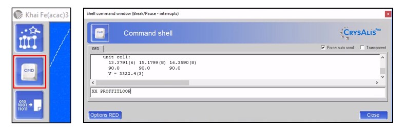
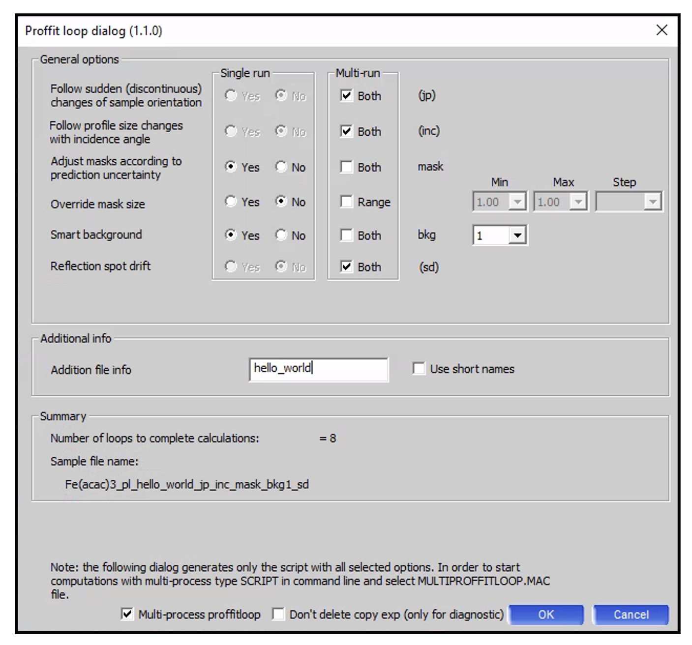
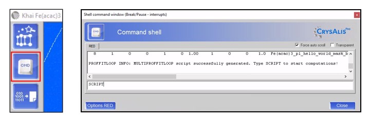
How do I use it? Open a *.par file in the offline version of CrysAlisPro, click on the power tool Command shell, and type “XX PROFFITLOOP” into the command line. | | Figure 1. Access the CrysAlisPro feature Proffitloop. | After pressing <Enter>, the [Proffit: CrysAlisPro data reduction assistant] wizard will appear, where a user selects default integration settings for all processing jobs. Proceed here as usual. While running Proffitloop, CrysAlisPro uses the same orientation matrix for all processing runs. Once all settings for the six steps are selected, press <Finish>. A new [Proffit loop dialog] GUI will appear, where some parameters can be changed. For more information on these parameters, please visit this link at the Rigaku X-ray Forum. | | | Figure 2. Proffitloop GUI, which is responsible for the script generation. | You can now choose which of the parameters will be used (Single run: Yes), or not be used (Single run: No), or tested for both options (Multi-run: Both) during multiple data reduction. Depending on the different parameter combinations, a number of loops will be generated and saved as *.proffitpars files (see Summary info box. In this case, Number of loops = 8). The files are saved under the following file name template: Experiment name (here: “Fe(acac)3”) “pl” is an abbreviation for proffitloop and indicates that the respective file was generated by the proffitloop dialog. If you tick the Use short names box, “pl” is removed from the file name. “hello_world” can be replaced with additional information given by the user in the edit box on the GUI. Please do not use any special characters. Use lowercase letters. Use underscores (“_”) instead of blank spaces. Parameters summary (here: “jp_inc_mask_bkg1_sd”; short names: “j_i_m_b1_s”). Information on which of the parameters were used for the respective data reduction.
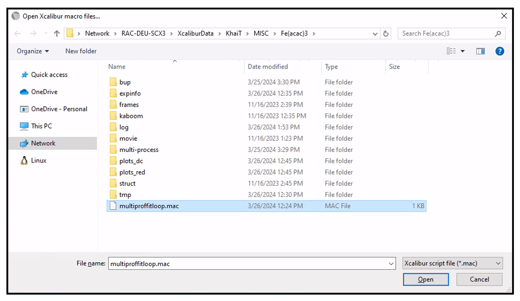
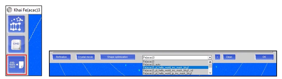
Selecting Multi-process proffitloop will increase the processing speed, and consequently decrease the processing time significantly, and thus, is highly recommended. After pressing <OK>, you can now choose the cores for subprocesses. Once you press <Run with sub processes>, please go back to power tool Command shell, type “SCRIPT” into the command line and open multiproffitloop.mac. The Proffitloop script will now run unattended for a few minutes. | | | | Figure 3. How to run the multi-process proffitloop. | After successful completion of the script, the files are stored in the experiment folder, and the results can be examined using the power tool Data finalization. The best parameter combination for data reduction for each dataset run can be evaluated by clicking on the power tool icon and scrolling through the data reduction output using the drop-down menu at the bottom of the window. By looking at the statistics printout tables (e.g. I/sigma, Rint values) for each processed dataset, you will be able to identify the best combination of reduction options to produce the best dataset. | | Figure 4. Results of each computation can be viewed in the DC RRP window. | Note: Threshold-based decisions may change depending on the outcome of the data reduction. The most obvious change that can happen is that different space groups or crystal systems are selected, giving drastically different values and making one result seem potentially worse than it is in reality. | | | | | | | | Review: Her Space, Her Time: How Trailblazing Women Scientists Decoded the Hidden Universe By Shohini Ghose ISBN 9780262048316
Shohini Ghose’s Her Space, Her Time: How Trailblazing Women Scientists Decoded the Hidden Universe shines a light on dozens of women whose contributions to the field of physics went unacknowledged in their own time—and, for many, even in ours. Some names are familiar, such as Lise Meitner and Marie Curie. But most of the names will be new for the average reader, although that is a statement about sexism and racism in historical and modern physics and not a reflection on the importance of these women’s contributions.
Each of Ghose’s seven chapters dives into the careers and biographies of these women scientists, albeit briefly. The women are grouped by the related natures of their research and discoveries, from space to radioactivity to subatomic particles. Since Her Space, Her Time is a short book, a little over 210 pages not counting the reference material in the back, Ghose does not take deep or detailed dives into these women or their research. But that works for Ghose, especially since one gets the sense that, in some of these cases, there just isn’t that much information about these women and their research, either publicly or readily available.
However, the most compelling part of the book is Ghose’s own interjected narrative. Each chapter has a smattering of anecdotes and asides where Ghose parallels the historical experience of a woman in her field with her own experience today (and in the recent past). Comparing her studies to that of another woman born in India 60 years prior, Ghose acknowledges that a lot happened in those 60 years—and yet some elements of their experiences were inevitably the same. It’s easy to read about the past and shrug it off as history—the present must surely be an improved time. But it’s another thing to have an author who is not afraid to share their personal experiences and root the reader in a contemporary reality where, yes, some things have changed for the better and yet there is still endless room for improvement.
Review by Jeanette S. Ferrara, MFA |
|
| | UPCOMING WEBINAR: TOPIQ | Illuminating the World of Sub-micron Crystal Structures with the XtaLAB Synergy-ED: a Review Wednesday, May 29, 2024
at 16:00 CET
Explore the diverse realm of sub-micron crystal structures through the lens of the XtaLAB Synergy-ED. This webinar delves into its applications across a wide spectrum of materials, including small molecules, MOFs, COFs, biological samples, minerals, and beyond. Highly beam-sensitive samples or measured under cryogenic conditions, the XtaLAB Synergy-ED offers exceptional capabilities for precise analysis. Discover how this instrument contributes to advancements in chemistry, materials science, and related fields. Register now > | | UPCOMING EVENTS:
American Crystallographic Association Annual Meeting, July 7-12, Denver, CO
Denver X-ray Conference, August 5-9, Denver, CO
European Crystallographic Meeting 34, August 26-31, Padova, Italy
Second meeting of the Latin American Crystallographic Association (LACA), October 23-27, Mérida, Mexico | | | | FOLLOW US ON TWITTER
To keep up to date on the latest news and events from Rigaku Oxford Diffraction, follow our Twitter feed. | | | JOIN US ON LINKEDIN
Our LinkedIn group shares information and fosters discussion about X-ray crystallography and SAXS topics. Connect with other research groups and receive updates on how they use these techniques in their own laboratories. You can also catch up on the latest newsletter or Rigaku Journal issue. We also hope that you will share information about your own research and laboratory groups. | | | RIGAKU X-RAY FORUM
At rigakuxrayforum.com you can find discussions about software, general crystallography issues and more. It’s also the place to download the latest version of Rigaku Oxford Diffraction’s CrysAlisPro software for single crystal data processing. | |
|
|