INTRODUCTION
Earlier this month, the Nobel Prizes were awarded for the work that provided the RNA vaccines used to combat COVID-19 (Medicine and Physiology), quantum dots (Chemistry) and attosecond atomic probes (Physics). The first two have impacted our lives in the form of vaccines, and brighter displays and biological probes, respectively. The latter work provides deep insight into electronic structure. All represent a lifetime of work in the researchers’ respective fields.
This month, we highlight our researcher of the month by e-interviewing Micheal Bodensteiner of the University of Regensburg. Fraser White provides a detailed look at Ewald 3D as the tip of the month. Our instrument in the spotlight is the popular XtaLAB Synergy-DW.
This month, Jeanette reviews The Deadly Rise of Anti-science: A Scientist’s Warning by Peter Hotez. I moved this book to the front of the queue, ahead of American Prometheus and Elon Musk, since the topic is so important: understanding how anti-scientists think is crucial to fighting anti-science.
I want to close with an observation I made while wandering around Freeport, Maine while on vacation. I saw a T-shirt with the word “humankind” and underneath “be both.” If we heed these words, the world will be a better place.
Be safe, Joe Ferrara | | | INTERVIEW WITH A RESEARCHER | | | Dr. Michael Bodensteiner
Michael has been the head of the department of X-ray Structure Analysis at the University of Regensburg since 2011. Prior to that he did his PhD in the group of Prof. Manfred Scheer on phosphanylalane chemistry.
In more recent years, Michael has become involved in Olexsys Ltd. and was appointed Managing Director of OlexSys GmbH in 2018. How would you describe your main research interest to a layman in one sentence?
I do fancy anomalous research with colorful X-rays.
What research project(s) or publications are you working on that you are most excited about right now?
I am still very excited to refine the anomalous dispersion parameters with Olex2 for every type of structure I get (IUCrJ 2022, 9, 604-609). It is really fascinating every time to get one step closer to the dream of one day enabling chemists to determine the oxidation state of an element with an in-house diffractometer.
What is the biggest challenge you are facing in your research just now?Currently, the most difficult thing for me is to try to be a good family man, service crystallographer, colleague, supervisor, lecturer and researcher at the same time during my much too short days.
Looking forward, where do you see the future of crystallography in the home lab leading?
I hope to see a wider range of X-ray diffractometers with more different wavelengths in the not-too-distant future. Copper K β radiation, for example, has served us very well in Regensburg for many years. I could also imagine that some X-ray spectroscopy techniques could be very well accommodated in a diffractometer cabinet.
Looking back, how has crystallography in the home lab changed for you and what have been the greatest advances you’ve seen since the start of your career? I started my scientific career as a young graduate student in 2006, when the Oxford Diffraction Gemini diffractometer had just been installed at our facility and, for the first time, experiments could routinely be measured in less than a day. The duration of the measurements reduced progressively as we got our SuperNovas and at the same time the quality of the data increased. The biggest leap in development for us was definitely the XtaLAB Synergy-DW with the HyPix Arc 150° detector, which was installed in October 2020 in our lab. I will never forget the faces of my colleagues when they saw the speed of the goniometer and that every few minutes a measurement was completed. I recently had the pleasure of measuring crystals on a XtaLAB Synergy-ED electron diffractometer that were invisible even in a very good microscope and I probably looked the same.
Anything else you’d like to share with our readers?
Stay curious!
| | | | Dr. Thomas Turner is a post-doctoral research associate in instrument design and flow systems at the School of Chemistry, University of Leeds. He completed his doctoral studies at the University of Leeds, where his research interests were focused on crystallization and the characterization of organic and pharmaceutical materials, in particular the study of nucleation using in-situ synchrotron X-ray scattering. Currently his research focus is on the development of a new EPSRC funded national facility, FlowXl, which utilizes state of the art in-situ X-ray diffraction combined with Raman spectroscopy to probe nucleation and crystallization processes of materials in flow. | | | | Thomas will be presenting a new TOPIQ webinar for us on November 15 at 4PM CEST entitled “Flow-Xl: A New UK Facility for the Analysis of Crystallisation in Flow Systems.”
| | | | | Dual-Wavelength Rotating Anode X-ray
Diffractometer with HPC X-ray Detector | | | | XtaLAB Synergy-DW VHF The introduction in 2004 of the Oxford Diffraction Gemini diffractometer, with two independent X-ray sources, was a watershed moment in crystallographic instrumentation. The groundbreaking design of the Gemini suddenly gave crystallographers the ability to easily switch between Cu and Mo wavelengths and greatly expanded the experimental flexibility available for analyzing single crystal samples. The XtaLAB Synergy-DW VHF is an evolution of that revolutionary idea which retains the flexibility of the dual wavelength capability but in addition adds the exceptional flux enhancement of a reliable, rotating anode X-ray source. It is the perfect diffractometer for a core facility where protein crystallography and small molecule crystallography are both practiced.
Configuration The XtaLAB Synergy-DW VHF diffractometer contains a PhotonJet-R X-ray source that is based on the proven, low-maintenance MicroMax-007 HF microfocus rotating anode X-ray generator. The target is constructed with two different X-ray source materials (the following combinations are available: Mo/Cu, Cu/Cr, Cu/Co, Cu/Ag, and Ag/Mo; only Mo and Cu optics are available with VHF style optics) and is coupled with an auto-switching dual-wavelength optic. Two wavelengths of X-ray radiation are available at the click of a button and switching between wavelengths takes only 5 minutes. Rounding out the XtaLAB Synergy-DW VHF configuration is the fast and efficient four-circle kappa goniometer which is coupled with Rigaku’s Hybrid Photon Counting (HPC) X-ray detector, the HyPix-6000HE (or optionally the curved, large theta coverage detectors, HyPix-Arc 100° or HyPix-Arc 150°.) which has essentially no readout noise, no dark noise and high dynamic range. All of this controlled by the CrysAlisPro diffraction software package with sophisticated algorithms to tie the hardware together to minimize the time it takes to measure and solve single crystal X-ray structures.
Proven Reliability The PhotonJet-R source was designed with reliability in mind. Clever Rigaku engineering makes filament changes easy, like swapping a printer cartridge, with no need to realign the source each time. Scheduled maintenance involves one annual visit from a Rigaku engineer, as with all XtaLAB Synergy diffractometers, and typically takes 1-2 days. With the anode exchange program, you get the benefit of rotating anode power with the convenience of sealed tubes.
Beam Conditioning Where overlapping peaks are a concern, e.g. large unit cells, proteins, twinned or incommensurate lattices, high beam divergence is undesirable. On PhotonJet sources, a software controlled, motorized variable beam slit is available as an option to alter divergence to adapt the source to your sample’s requirements. For those samples where intensity matters most, the slit can be fully opened giving the highest flux. For those where peak sharpness and overlap are factors, the beam can be limited to a divergence anywhere between 1 to 10 mrad.
CrysAlisPro The XtaLAB Synergy-DW VHF comes complete with CrysAlisPro, our user-inspired data collection and data processing software for single crystal analysis. Designed around an easy-to-use graphical user interface, CrysAlisPro can be operated under fully automatic, semi-automatic or manual control. CrysAlisPro combines automated crystal screening, the fastest and most accurate strategy software available, concurrent data reduction and automatic small molecule structure solution. CrysAlisPro can operate either in a protein or small molecule dedicated workflow. Popular third-party protein data processing packages can easily process diffraction data if desired. Visual feedback is provided for each step with clear, color-coded guidance so that both novices and experts can collect high-quality data in the shortest time possible.
AutoChem AutoChem is the ultimate productivity tool for small molecule chemists, offering fast, fully automatic structure solution and refinement during data collection. Developed in collaboration with OlexSys Ltd (Durham University, UK), AutoChem works in conjunction with Olex² where more advanced structure solution and refinement functionality exists. AutoChem is seamlessly integrated within CrysAlisPro, and forms an integral part of our ‘What is this?’ feature. The ‘What is this?’ feature gives you structures quickly and ensures you are not wasting time collecting full datasets on known samples or starting materials. It is an alternative pre-experiment option, which is used to plan your full data collections. | | | | Ewald3D
What is it? Ewald3D is a three-dimensional diffraction viewer that can be used to display real intensity from diffraction images. It is similar to a reciprocal lattice viewer, but with experimental intensities.
Why should I use it? When you view the diffraction pattern in 3D, it allows you to more easily spot issues with your sample or instrument setup. The Ewald3D view is unbiased by unit cell; therefore, even very weak intensity away from lattice positions can be spotted, leading to a better understanding of the crystallinity of your sample. It can help you spot issues like twinning, incommensurate behavior, diffuse scatter and other issues arising from non-Bragg intensity.
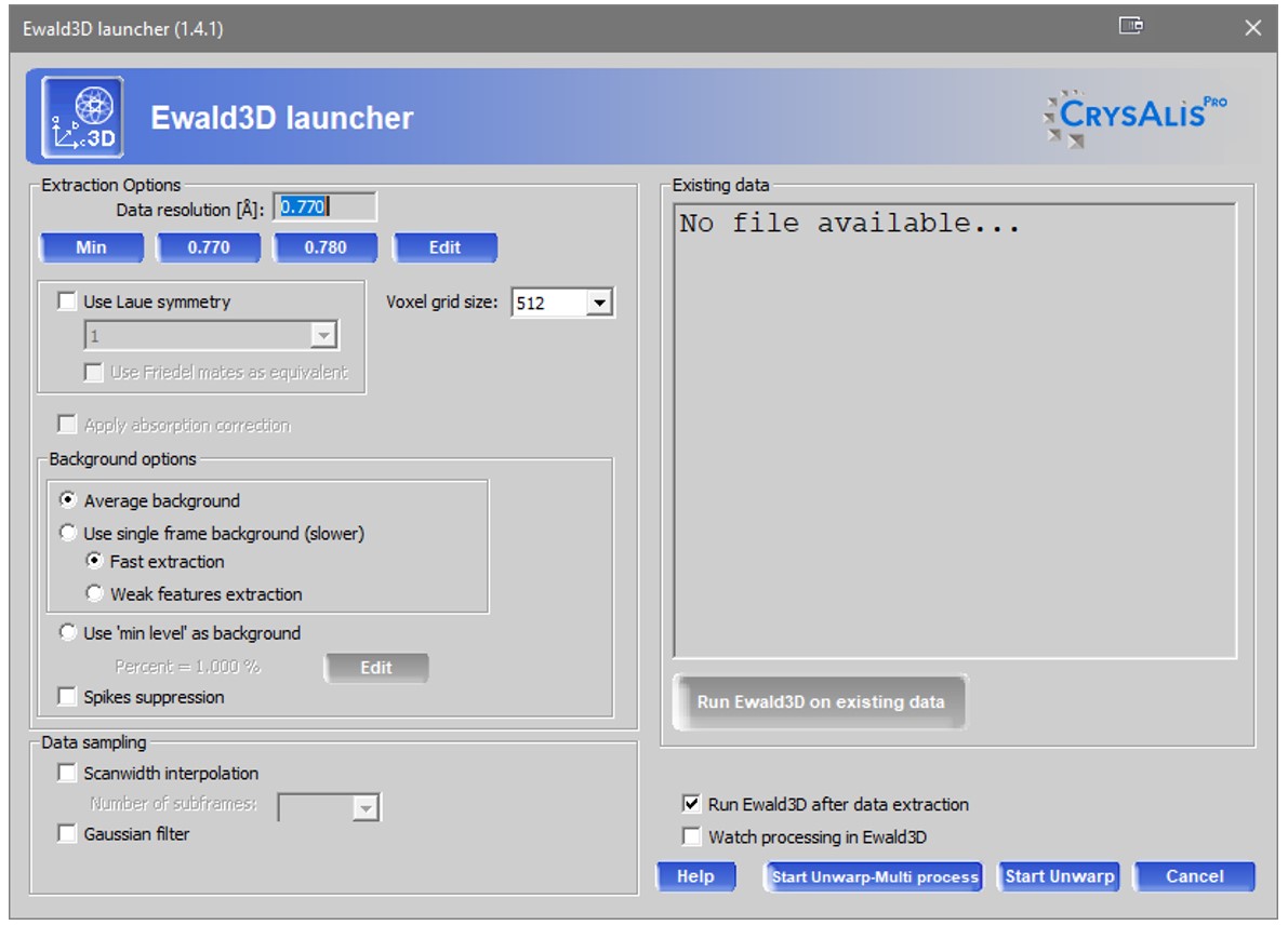
How do I use it? Ewald3D is accessed either during live experiments (depending on hardware) or via the lattice viewer at the conclusion of the experiment. The location of the launch icon is shown below. | | | Figure 1. The location of the launch icon. | | You have the several options to manipulate the dataset generation: Voxel size – larger voxels combine intensities from a larger number of pixels for weaker data; smaller voxels offer finer spatial resolution. Symmetry – use symmetry to complete/average intensities, giving a more complete view for high-symmetry species where a sphere of data was not collected. Background subtraction – you have some control over how background is evaluated, depending on how weak the features you are interested in are.
| | Figure 2. Ewald3D launcher. | | You can watch data extraction proceeding within Ewald 3D if desired, and Ewald 3D can even be used live during an experiment depending on your hardware.
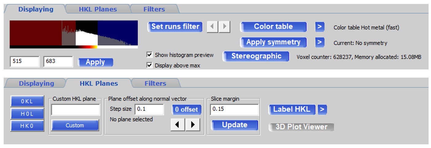
View types Data can be viewed in orthogonal or stereographic space, with custom intensity ranges, specific runs and with different color tables. | | | You can also view slices through the 3D plot similar to more traditional “unwarp” or simulated precession images. The advantage here is that you can interactively step through 3D space in increments without having to generate each slice one by one as with unwarp images.
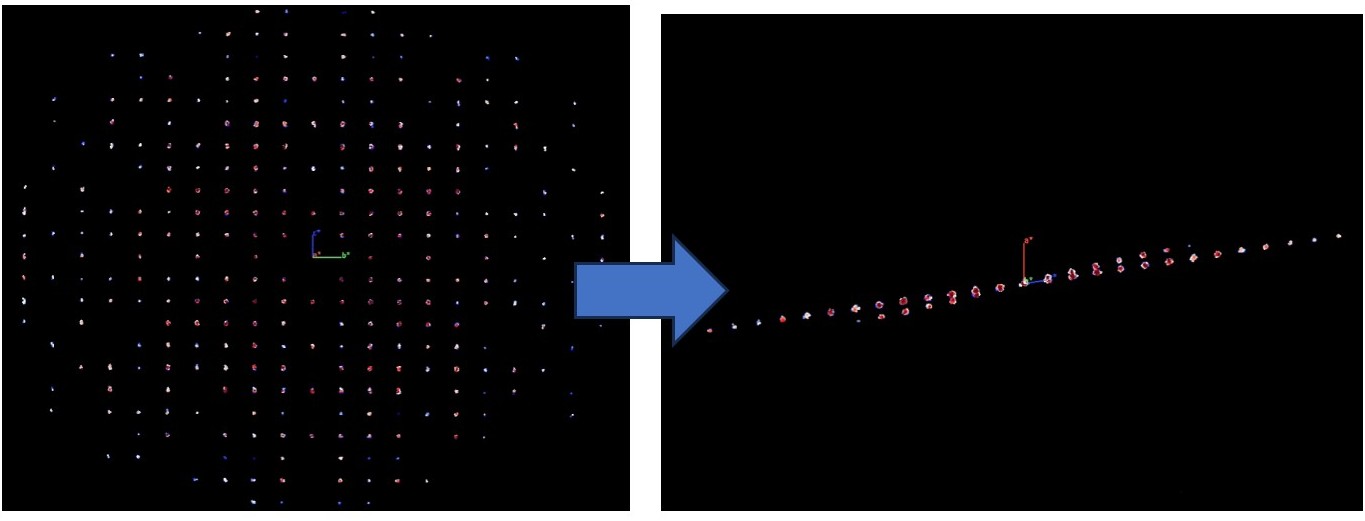
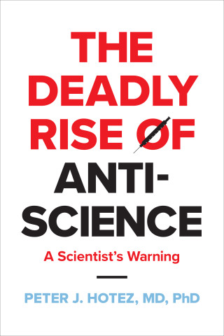
Example A simple twin with slight rotation between domains can be hard to spot. In some views, the twin is not obvious. Simply rotating the 3D diffraction space easily reveals the twin, when Ewald 3D is used live during data collection, the twin can be spotted quickly and easily without requiring unwarps, processing or twin indexing. | | Figure 4. A twin with slight rotation between domains. | | | | | | | Review: The Deadly Rise of Anti-Science: A Scientist’s Warning By Peter J. Hotez ISBN 9781421447223
Disclosure: The reviewer has interviewed Dr. Hotez multiple times, with Dr. Hotez serving as an expert source while researching tropical infectious diseases.
Peter Hotez’s The Deadly Rise of Anti-Science: A Scientist’s Warning is a chilling dissertation and gut-wrenching glimpse into navigating the COVID-19 pandemic as a leading vaccine researcher. Hotez is a professor of pediatrics and molecular virology at Baylor College of Medicine, as well as co-director of the Texas Children’s Center for Vaccine Development. As an MD-PhD, Hotez was uniquely qualified and positioned at the start of the COVID-19 pandemic to make an impact on vaccine development and adoption. Indeed, he and his research partner, Dr. Maria Elena Bottazi, were nominated for a Nobel Peace Prize for their efforts to develop and distribute an affordable COVID-19 vaccine to underdeveloped countries around the world.
Hotez became more of a household name during the pandemic due to his frequent appearances on various television news programs promoting the importance of safe COVID-19 practices and staying vaccinated. Sadly, this also put Hotez–and his family–on the radar of various anti-vaccination organizations. The most disturbing part of Hotez’s book isn’t necessarily his description of the rise of anti-science (which is in and of itself disturbing), it’s the excerpts he includes from various intimidating emails he has received throughout the pandemic. The hatred in these emailed death threats is startling and unnerving. If much of The Deadly Rise of Anti-Science feels like Hotez justifying himself and his pursuit of science as a means to reduce the toll of human lives lost during not just this pandemic but any pandemic, one can understand why, given the volume of vociferous and violent feedback he’s received.
Hotez defines anti-science as “the rejection of mainstream scientific views and methods or their replacement with unproven or deliberately misleading theories, often for nefarious and political gains.” He draws an interesting parallel between how certain political groups in this country disseminate misinformation and seek to discredit reputable researchers with how it was done by the Communist government in the Soviet Union. Given the disdain certain political parties in America have for the concept of anything remotely related to communism, it’s certainly intriguing to know that the same metaphorical weapons of misinformation fill their arsenals.
The audience who needs to read The Deadly Rise of Anti-Science probably isn’t the audience that will. Regardless, it provides compelling insight as to why science, especially medical science, has become widely (and violently) politicized in the United States.
Review by Jeanette S. Ferrara, MFA |
|
| | | | UPCOMING EVENTS: SERMACS 2023, Durham, NC, October 25-28, 2023.
31st Protein Structure Determination in Industry, Cambridge, UK, November 12-14, 2023.
Rayons X et Matière 2023, Bordeaux, France, November 21-24, 2023. | | USEFUL LINKS
All ACA Members are invited to submit videos to be eligible for a $500 (USD) prize. Videos will be evaluated by the Young Scientists Interest Group, and the Education Committee will award the winner. The first 10 submissions before the October 31, 2023 deadline will be automatically eligible for a $100 (USD) prize. Submit a video>
Please note: this is open to everyone, with a small cash donation to the bank of yourself! The video does not need to be a full-lecture. Five to ten-minute videos are encouraged!
| | We now have a full-scale war in the Middle East, earthquakes In Afghanistan an ongoing war in Ukraine, and humanitarian crises elsewhere. With some much suffering, perhaps it is a good time to donate to larger relief organizations like the International Red Cross and Red Crescent Movement, so they can distribute relief as needed. | | FOLLOW US ON TWITTER
To keep up to date on the latest news and events from Rigaku Oxford Diffraction, follow our Twitter feed. | | | JOIN US ON LINKEDIN
Our LinkedIn group shares information and fosters discussion about X-ray crystallography and SAXS topics. Connect with other research groups and receive updates on how they use these techniques in their own laboratories. You can also catch up on the latest newsletter or Rigaku Journal issue. We also hope that you will share information about your own research and laboratory groups. | | | RIGAKU X-RAY FORUM
At rigakuxrayforum.com you can find discussions about software, general crystallography issues and more. It’s also the place to download the latest version of Rigaku Oxford Diffraction’s CrysAlisPro software for single crystal data processing. | |
|
|